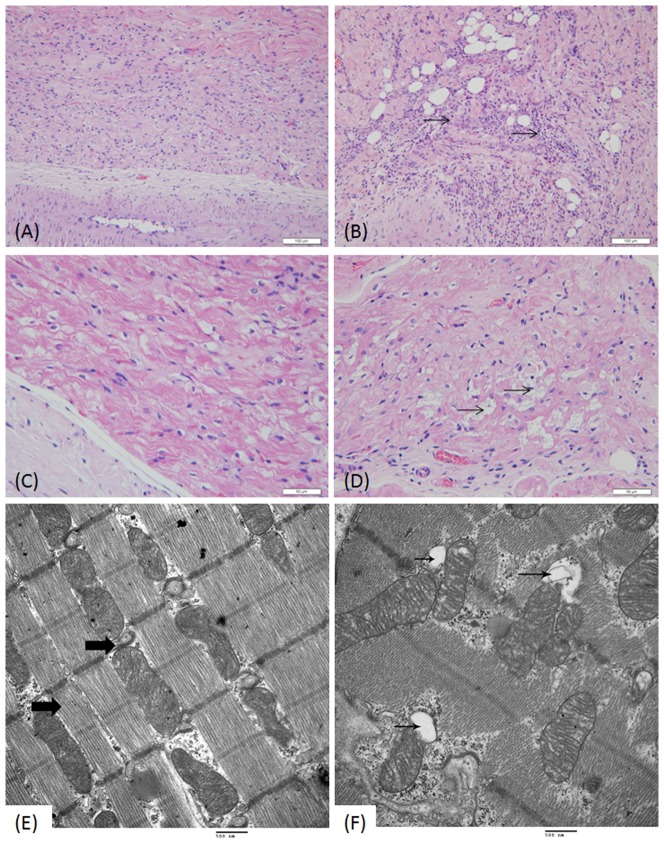
Figure 3. Comparison of the CCS morphology in the heart of dog receiving vehicle only (A, C, and E) and ART (6 mg kg−1) (B, D, and F).
Hematoxylin and eosin staining show inflammation and fatty infiltration in the sinoatrial node (SAN) area of the cardiac conduction system (black arrows, B) and vacuolar degeneration of some pacemaker cells in the AVN (black arrows, D). Ultra-structural mitochondria in myocardial cells show mitochondrial bending, distortion, and swollen vacuoles (black arrows, F). (Original magnification of A and B×200, C and D×400, TEM image, 25,000×.)

