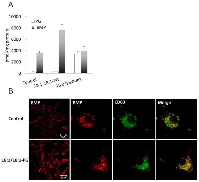
Figure 1.
Quantification of BMP and cellular localization. (A) BMP and PG from control cells, 18:1/18:1-PG and 16:0/16:0-PG supplemented cells were quantified by LC-MS as described in Materials and Methods. Data are the mean ± SD of 3 independent determinations. (B) Control and BMP-enriched cells were fixed and stained with Alexa 546-conjugated anti-BMP antibody (6C4) and with Alexa 488-conjugated anti-CD63 antibody. Bar, 10 μm.

