Figure 6.
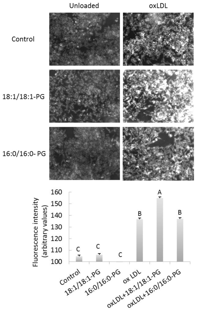
BMP accumulation enhances foam cell formation. Control, BMP and PG-enriched cells were incubated in basal conditions (unloaded) or exposed to 50 μg/mL oxidized LDL for 24h to induce foam cell formation. Cells were fixed and stained with Nile Red. (A) Stained cells were observed through fluorescent microscope and images show representative Nile Red-stained cells. Bar, 25 μm. (B) Quantification of fluorescence was determined using cell^Fsoftware and values represent the means ± SD of four fields. A, B, C, significantly different groups (multiple means comparisons by ANOVA and Tukey-Kramer method with α= 0.05).

