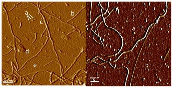
Figure 1. Components of aggregating lysozyme.
Left: AFM image showing various structures seen in aggregating lysozyme on mica substrate: (a) colloidal spheres, (b) primary fibers, (c) compound fibers, (d) amorphous aggregates, (e) a continuous layer of protein monomers bound to substrate. Right: After 1∶1000 dilution the continuous protein layer is no longer present. Colloidal spheres are still seen (a), as are numerous discrete particles (b) with a height of  nm (n = 20), probably representing individual lysozyme monomers.

