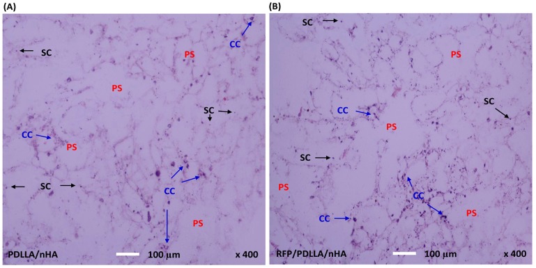
Figure 5. Histological evaluation of MC3T3-E1 cells.
After 7 days of co-culture with MC3T3-E1 cells, the horizontal composites sections with a thickness of 5 µm were stained with hematoxylin-eosin and observed by a Leica light microscopy (400x magnifications). MC3T3-E1 cells proliferated within the two kinds of composites, and formed numerous cell colonies (CC). Cells stably adhered to the walls of the inner holes and were randomly distributed along the walls. A: PDLLA/nHA, B: RFP/PDLLA/nHA; PS: porous structure;SC: single cell. [Scale bar, 100 µm].

