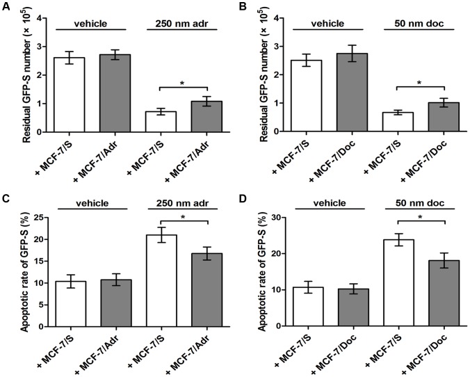
Figure 1. Effects of cell co-cultures.
(A) Number of residual GFP-S was evaluated after cell mixture was treated with vehicle or 250 nm adr for 24 h. (B) Number of residual GFP-S was evaluated after cell mixture was treated with vehicle or 50 nm doc for 24 h. (C) Apoptotic rate of GFP-S was determined after cell mixture was treated with vehicle or 250 nm adr for 24 h. (D) Apoptotic rate of GFP-S was determined after cell mixture was treated with vehicle or 50 nm doc for 24 h. Data are expressed as the mean ± SD, n = 3: * P<0.05, + MCF-7/Adr vs. + MCF-7/S or + MCF-7/Doc vs. + MCF-7/S.

