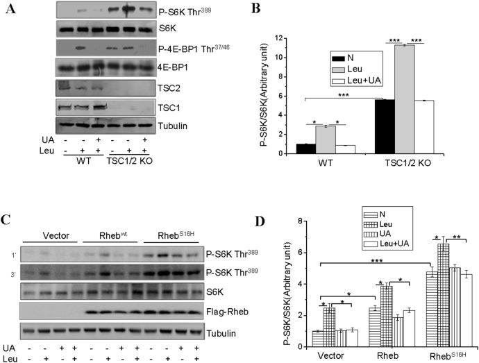
Figure 2. UA inhibits leucine-stimulated mTOR activation in a TSC1/2 and Rheb-independent manner.
(A), Serum-starved TSC1/2+/+ (WT) and TSC1/2−/− (KO) MEF cells were pretreated with or without 50 µM for 60 min and then treated with or without 10 mM leucine (Leu) for 60 min. The phosphorylation and protein levels of S6K, 4E-BP1 and the protein levels of TSC1 and TSC2 in cell lysates were determined by Western blot with the indicated antibodies. (B), S6K phosphorylation shown in (A) was semi-quantified and normalized to S6K protein levels. (C), serum-starved C2C12 cells transiently expressing Rheb or RhebS16H were treated with or without 50 µM UA for 1 hour, followed with or without 10 mM leucine (Leu) for 60 min. The phosphorylation and protein levels of S6K, and the protein levels of FLAG-Rheb in cell lysates were determined by Western blot using indicated antibodies. (D), S6K phosphorylation in (C) was semi-quantified using the NIH Image J Program and normalized with S6K protein levels. Differences between groups were examined for statistical significance using ANOVA. Data are presented as mean ± S.E.M. from three independent experiments. *, P<0.05, **, P<0.01, and ***, p<0.001. N, no addition.

