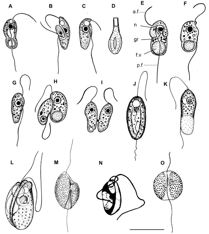
Figure 7. Drawings of colponemids.
(a–d) Colponema edaphicum (from Mylnikov and Tikhonenkov [43]), (e–i) C. marisrubri (from Mylnikov and Tikhonenkov [25]), (j–m) C. loxodes ((j, k) from Zhukov and Mylnikov [41]; l) from Chadefaud [42]; m) from Lemmermann [38], n) C. symmetricum (from Sandon [39]), o) C. globosum (from De Faria et al. [51]). a.f – anterior flagellum, f.v – food vacuole, gr – longitudinal groove, n – nucleus, p.f – posterior flagellum. Scales: (a–c), (e–i) –10 µm; d) –1; (j–m) –20 µm; (n, o) –15 µm.

