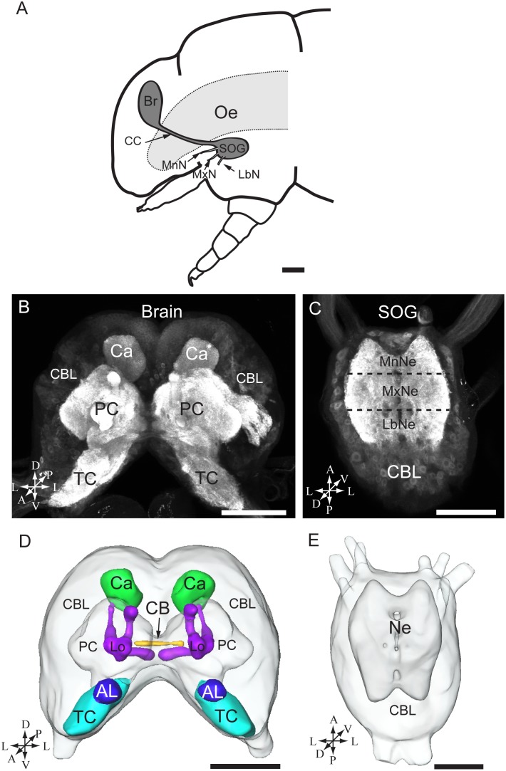
Figure 2. The brain and the suboesophageal ganglion of the fifth instar larvae of H. armigera.
(A) Diagram of the larval head including the brain (Br) and the suboesophageal ganglion (SOG). (B) Confocal image of the brain showing the cell body layer and the central neuropils. (C) Confocal image of the SOG showing the cell body layer (CBL) and the three fused neuromeres, from anterior to posterior: the mandibular neuromere (MnNe), the maxillary neuromere (MxNe), and the labial neuromere (LbNe). (D) 3D reconstruction of the whole brain. (E) 3D reconstruction of the SOG. A anterior, D dorsal, L lateral, M medial, P posterior, V ventral. Oe oesophagus, CC circumoesophageal connectives, MxN maxillary nerve, LbN labial nerve, MnN mandibular nerve, TC tritocerebrum, PC protocerebrum, Ca calyces of the mushroom body, AL antennal lobe, CB central body, Lo lobles, Ne neuromere. Scale bars: 250 µm in A, 100 µm in B, C, D, and E.

