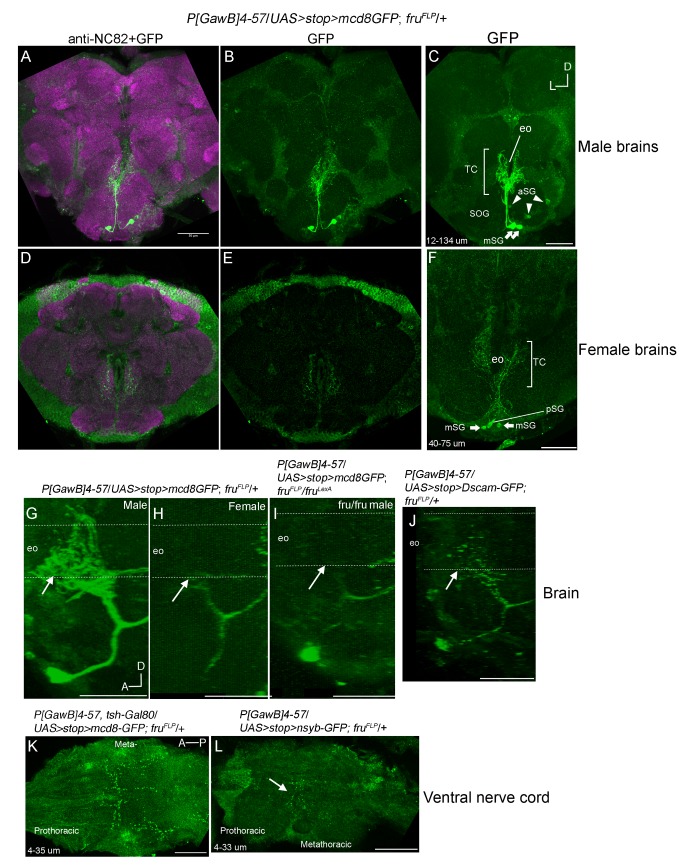
Figure 4. Visualization of fruM(+) and P[GawB]4-57 intersection revealed a sexually dimorphic arborization in the tritocerebrum.
A–F) Anterior-posterior, G–J) sagittal, and K–L) dorsal-ventral confocal projections. Unless otherwise stated, images are from males. A–I) Z-projections showing GFP fluorescence from male P[GawB]4-57/UAS>stop>mCD8-GFP; fruFLP/+ brains. A–F) Merged images showing anti-NC82 and GFP expressions in male and female brains. In all brains two GFP(+) cell bodies, in the ventral medial SOG (mSG, arrows) project to and make extensive arborizations in the tritocerebrum. C) In 7/14 brains, 3 cell bodies (aSG, arrowheads) fluoresced at depths of 17–20 µm without detectable neurites. D–F versus A–C) Z-projection showing the weaker tritocerebral aborizations from the two mSG neurons in female P[GawB]4-57/UAS>stop>mCD8-GFP; fruFLP/+ brains. F) pSG marks one posterior GFP(+) neuron. G–I), sagittal reconstructions of mSG projections in a G) P[GawB]4-57/UAS>stop>mCD8-GFP; fruFLP/+ male, H) female, and I) a P[GawB]4-57/UAS>stop>mCD8-GFP; fruFLP/fruLexA male mutant brain. Dashed lines mark the path of the esophagus. In fru +/fru - males the tritocerebral arbors are significantly larger and fluoresce brighter compared to fru +/fru - females or fru mutant males (arrows). J) Expression of the dendritic marker UAS>stop>Dscam17-1-GFP in mSG∩4-57 neurons colocalized with the tritocerebral arbors and anterior to medial tracts. L) Presynaptic marker, UAS>stop>nsyb-GFP was expressed mainly in prothoracic/metathoracic boundary. K) The presence of tsh-Gal80 repressed expression from fruM(+) ventral nerve highlighting the descending projections from mSG and pSG cells. Scale bars = 50 µm. eo = esophageal foramen, TC = tritocerebrm, and SOG = subesophageal ganglion.

