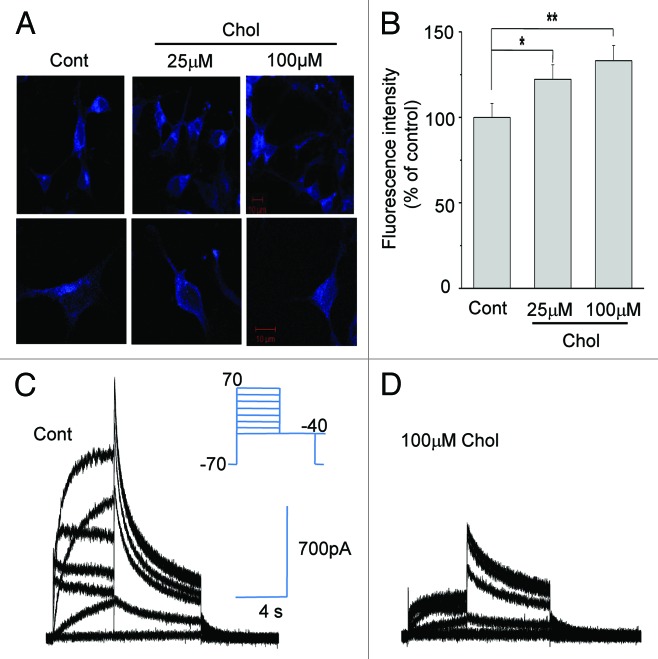
Figure 1. Increasing membrane cholesterol levels inhibits HERG K+ currents. (A and B) Incubating HERG-transfected HEK293 cells with MβCD-cholesterol increased cholesterol levels in the plasma membrane. Cells were incubated with 25 μM or 100 μM MβCD-cholesterol for 1 h at 37 °C. Filipin staining was performed for 30 min at room temperature after cholesterol enrichment. Typical fluorescent images are shown (A). The scale bars represent 10 μm. Fluorescent intensities from plasma membrane were quantified as described in Materials and Methods (B; n = 38). *p < 0.05; **p < 0.01 (C and D) HERG K+ currents were inhibited by cholesterol enrichment. Cells were incubated with 100 μM MβCD-cholesterol for 1 h before recordings. Representative families of HERG K+ currents from control cells (C) and from cholesterol-enriched cells (D) were recorded using the voltage protocol shown in the inset. Depolarizing steps were applied from a holding potential of −70 mV to between −60 and +50 mV for 4 sec, followed by a step to −40 mV to elicit currents.

An official website of the United States government
Here's how you know
Official websites use .gov
A
.gov website belongs to an official
government organization in the United States.
Secure .gov websites use HTTPS
A lock (
) or https:// means you've safely
connected to the .gov website. Share sensitive
information only on official, secure websites.
