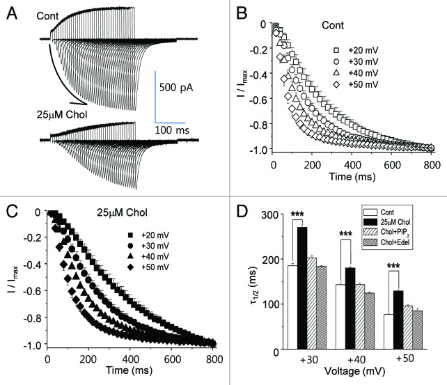
Figure 5. Activation kinetics of HERG current are slowed by mild cholesterol enrichment due to the activation of PLC. HERG current was activated by varying the duration of the depolarizing pulse at different testing voltages. The peak inward tail current was recorded following repolarization to −110 mV. (A) A typical recording at +40 mV testing voltage is shown from control cells and from cells incubated with 25 μM MβCD-cholesterol for 1–2 h at 37 °C before recordings. The arrow in the figure represents the accumulation of peak inward tail currents elicited by increasing the duration of the depolarizing pulses. (B and C) Summary of activation kinetic analysis as determined by time constants fitted to depolarization-induced outward currents shows that mild cholesterol enrichment decelerated channel activation. Each symbol represents time constants from different testing voltages (+20, +30, +40 or +50 mV; n = 6). (D) The half times to maximal rise (τ1/2) were obtained by fitting the curves at (B), and (C) with single exponential decays. τ1/2 obtained at different testing voltages (+20, +30 or +40 mV) show that the mild cholesterol enrichment decelerated channel activation. In some recordings, cells were treated with MβCD-cholesterol along with the PLC inhibitor, 5 μM edelfosine (Chol+Edel; n = 7), or 25 μM PtdIns(4,5)P2 was included in the pipette solution [Chol+PtdIns(4,5)P2; n = 6]. ***p < 0.001.

An official website of the United States government
Here's how you know
Official websites use .gov
A
.gov website belongs to an official
government organization in the United States.
Secure .gov websites use HTTPS
A lock (
) or https:// means you've safely
connected to the .gov website. Share sensitive
information only on official, secure websites.
