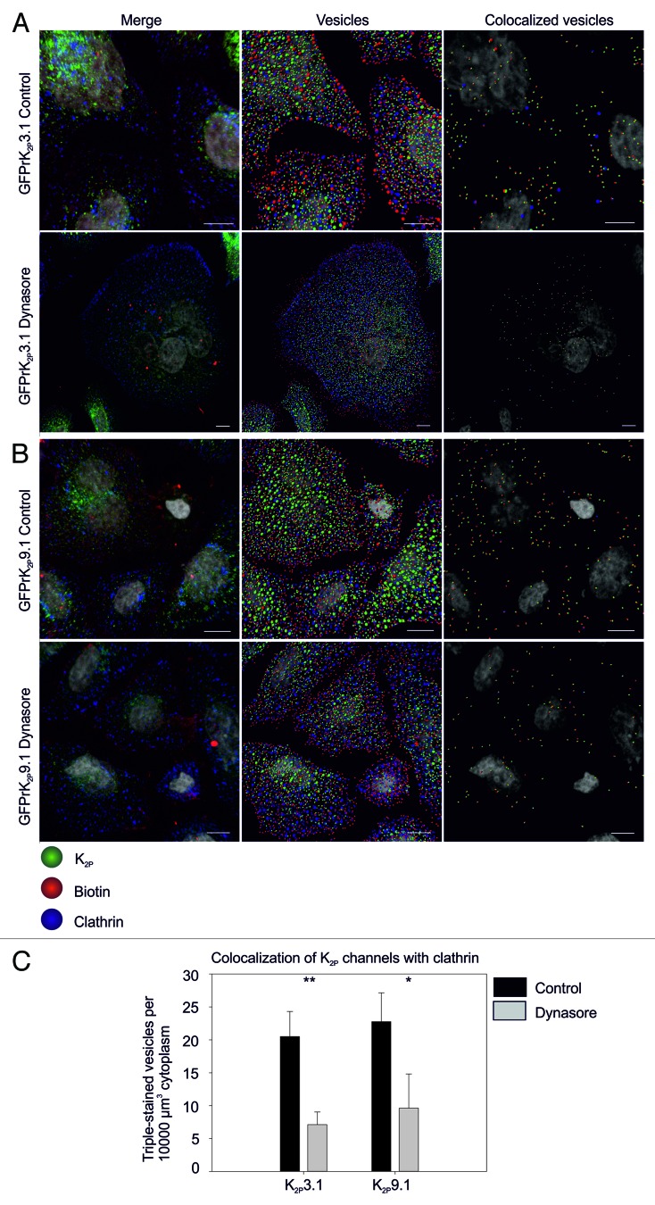
Figure 4. Dynamin-dependent endocytosis of GFPrK2P3.1 and GFPrK2P9.1. HeLa cells transiently expressing GFPrK2P3.1 [green, (A)] or GFPrK2P9.1 (green, (B)] were incubated with DMSO vehicle (Control) or the dynamin inhibitor, Dynasore (as indicated) before surface biotinylation on ice and warming to 37 °C for different periods of time. Images show cells stripped to remove surface biotin and fixed after a 30-min incubation period, before staining to detect biotin (red) and clathrin (blue). Merge: whole cell projection. Vesicles: image analyzed using Imaris to detect 0.6 μm and 1.2 μm spots. Colocalized vesicles: only the channel-biotin or channel-biotin-clathrin triple-colocalized 0.6 μm and 1.2 μm spots shown. Scale bars: 10 μm. (C) Images were analyzed by Imaris to calculate the number of triple-colocalized (channel, biotin and clathrin) 0.6 μm vesicles per unit volume of cytoplasm. Numbers are the mean and standard error from n = 3 fields of view. Inhibition by Dynasore is significant for GFPrK2P3.1 at p < 0.01; GFPrK2P9.1 at p < 0.05.

An official website of the United States government
Here's how you know
Official websites use .gov
A
.gov website belongs to an official
government organization in the United States.
Secure .gov websites use HTTPS
A lock (
) or https:// means you've safely
connected to the .gov website. Share sensitive
information only on official, secure websites.
