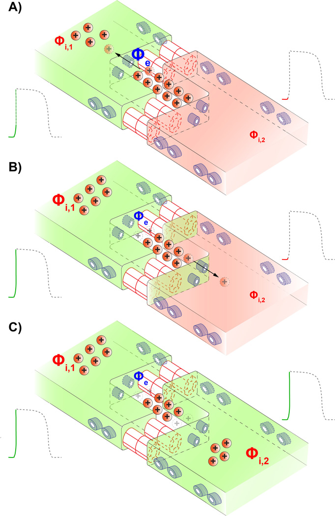
Figure 1.
Schematic cartoon illustrating the mechanism of ephaptic coupling. A) Sodium channels (shown in blue) on the depolarized myocyte's membrane activate, withdrawing positively charged sodium ions (Na+) from the restricted extracellular cleft at the intercalated disk. This raises the intracellular potential (Φi,1) of the first myocyte. B) Concomitantly, the depletion of positive charge from the restricted extracellular cleft lowers the local extracellular potential (Φe). There is a resultant increase in the transmembrane potential across the membrane of the second myocyte which is defined as the difference between its intracellular potential (Φi,2) and the extracellular potential (Φe). In turn sodium channels located at or near the intercalated disk of the second myocyte activate. C) Entry of sodium ions into the second myocyte via its sodium channels further depolarize it, triggering an action potential. Thus activation is communicated 'ephaptically' from cell-to-cell without the direct transfer of ions between them.

