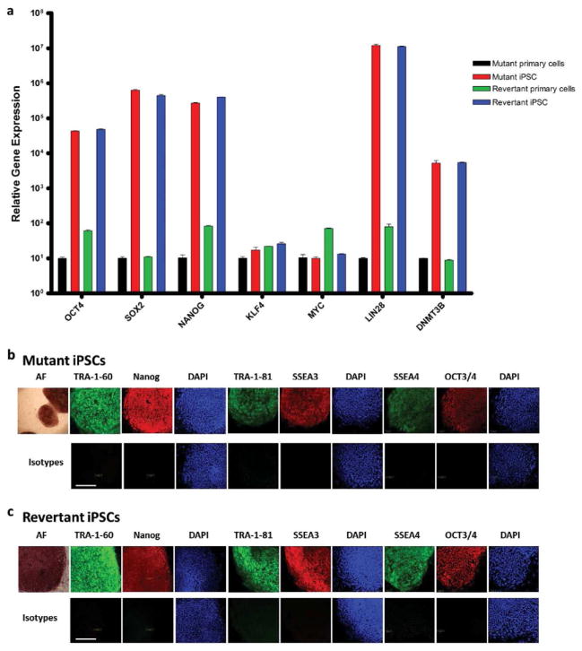
Figure 2. Revertant iPSCs from RDEB skin.
(a) Persistent mRNA expression of OCT4, SOX2, NANOG, LIN28, and DNMT3b, and transient mRNA expression of c-MYC and KLF4, both consistent with fully reprogrammed mutant and revertant iPSCs. (b) Protein expression in mutant iPSCs of embryonic stem cell surface markers TRA-1-60, TRA-1-81, SSEA3, SSEA4, alkaline phosphatase (AF), and transcription factors Nanog and OCT3/4. (c) Same embryonic stem cell protein expression panel in revertant iPSCs. Nuclei are stained with DAPI (blue). Lower panels show corresponding isotype controls. Scale bar = 50 μm.

