
Figure 4.
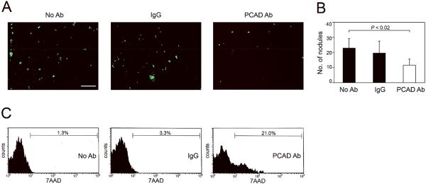
Blocking P-cadherin increases anoikis and decreases EOC cell attachment in vivo. (A,B) Female nude mice were injected i.p. with equivalent numbers of GFP-expressing control (non-targeting) SKOV3ip cells together with neutralizing Ab to mouse P-cadherin, control IgG or with no Ab. Mice were sacrificed at 10 days thereafter. (A) Tumor implants that were attached to the peritoneal cavity wall were viewed under a fluorescence stereoscope. Bar, 1 mm. (B) Numbers of tumor implants on the peritoneal cavity wall were counted in five random 25mm2 fields per mouse. (C) GFP-expressing control SKOV3ip cells were pre-incubated with neutralizing Ab to human P-cadherin, control IgG or with no Ab, and then injected i.p. into mice. Mice were sacrificed at 3 days thereafter. Floating cells in the peritoneal cavity were collected and stained with 7AAD. Cell death within the gated population of GFP+ tumor cells was evaluated by flow cytometric analysis of 7AAD staining.