
Figure 3.
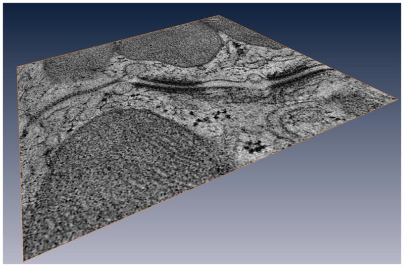
Vesicular activity at the intercalated disc. Tomographic electron microscopy image of an intercalated disc region. Notice the vesicular activity in the space between the desmosomes and the gap junction, as well as in the intercellular space. From67 with permission.