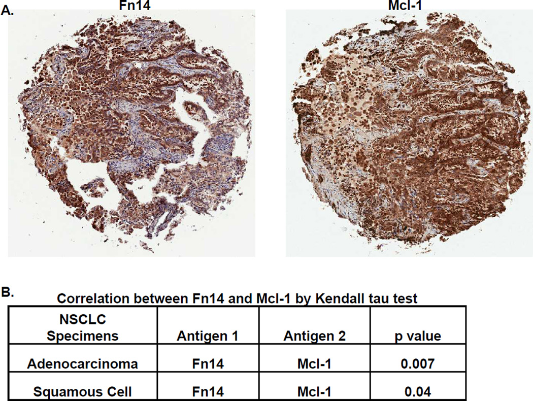
Figure 1. Mcl-1 expression in human NSCLC specimens correlates with Fn14 expression.
(A) Mcl-1 and Fn14 staining on representative samples from the same patient with lung adenocarcinoma (5× objective, Aperio GL Scanner). Tumor-cell specific Fn14 and Mcl-1 staining in each of the tumor punches was scored by a board-certified pathologist; a score of zero indicates staining level equal to adjacent non-tumor cells. A non-zero score indicates increased staining (1= minimum, 2= moderate, 3= strong positive). (B) A total of 290 samples were scored for Mcl-1 and Fn14 expression and the correlation between the two stains was analyzed using Kendall’s tau test.

