
Figure 3.
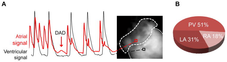
Anatomical origin of DADs in intact Casq2−/− atria. A) Representative example of a delayed afterdepolarizations (DAD). The red optical voltage record originating from the atria shows a deflection in the post pacing pause consistent with a DAD. Only deflections greater than 10% atrial of action potential amplitude were considered as DADs. No influence from ventricular fluorescence (black signal) is present during the atrial DAD. B) Anatomical distribution of DADs in Casq2−/− atria: The majority of the DADs occurred in the pulmonary vein region.