Figure 5.
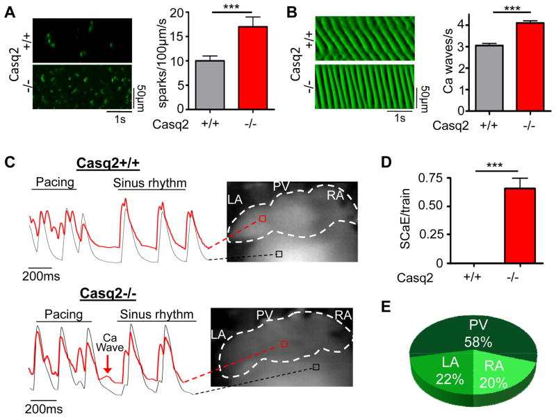
Ca handling is impaired in isolated atrial myocytes (A, B) and intact atria (C–E) of Casq2−/− hearts. A and B left panels: Representative line scans of Ca sparks (A) and Ca waves (B) obtained in permeabilized atrial myocytes. Right panels: averaged data. N=35–45 cells per group, ***P<0.001. C) Examples of fluorescent Ca signals obtained from Ca maps of Casq2+/+ and Casq2−/− hearts. Casq2−/− atria frequently exhibit spontaneous Ca elevations (SCaE) after the rapid pacing train that are absent in Casq2+/+ atria (D). Similar to DADs, SCaE were observed only in some regions of the atria. E) Anatomical distribution of SCaE in intact atria.

