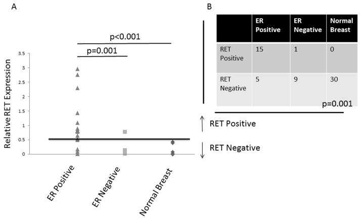
FIGURE 5. RET Expression is Associated with ERα Positive Breast Cancers.
A. RNA extracted from primary human breast cancers and patient matched normal breast tissue demonstrates significantly higher mean RET expression in ERα-positive tumors compared to ERα-negative tumors and normal breast tissue. The data represent values from 20 ERα-positive, 10 ERα-negative tumors, and the 30 samples of matched normal breast tissue, which are overlaid for some data points. The p values were calculated using the Student’s t-test. B. When a cutoff of 0.5 fold RET expression relative to MCF-7 is used, significantly more ERα-positive tumors are RET positive compared to ERα-negative and normal breast tissue, p=0.001. The p value was calculated using the analysis of variation (ANOVA).

