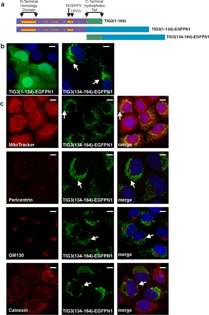
Fig. 2.
Role of the C- and N-terminal domains in guiding subcellular localization. A Schematic of TIG3-EGFPN1 fusion proteins. The proteins are as described in Fig. 1A, and the blue rectangle designates EGFPN1. B Subcellular localization of TIG31–134-EGFPN1 and TIG3134–164-EGFPN1 in SCC-13 cells. SCC-13 cells were electroporated with 3 µg of the indicated plasmid and after 24 h EGFPN1 fluorescence was detected by confocal microscopy. The arrows indicate the novel subcellular distribution of TIG3134–164-EGPN1. No signal was observed in non-electroporated cells (not shown). C TIG3134–164-EGFPN1 localizes at the mitochondria. Cells were electroporated with 3 µg of TIG3134–164-EGFPN1 and after 24 h the cells were fixed and stained with anti-pericentrin (centrosome), GM130 (Golgi), or calnexin (ER), or incubated with MitoTracker (mitochondria) (red). The images were then visualized by confocal microscopy. The merged images are the composite of the green and red channels and yellow indicates co-localization. Arrows indicate perinuclear TIG3 localization. Red, green (EGFPN1) and merged channels are shown. Bars = 10 µm.

