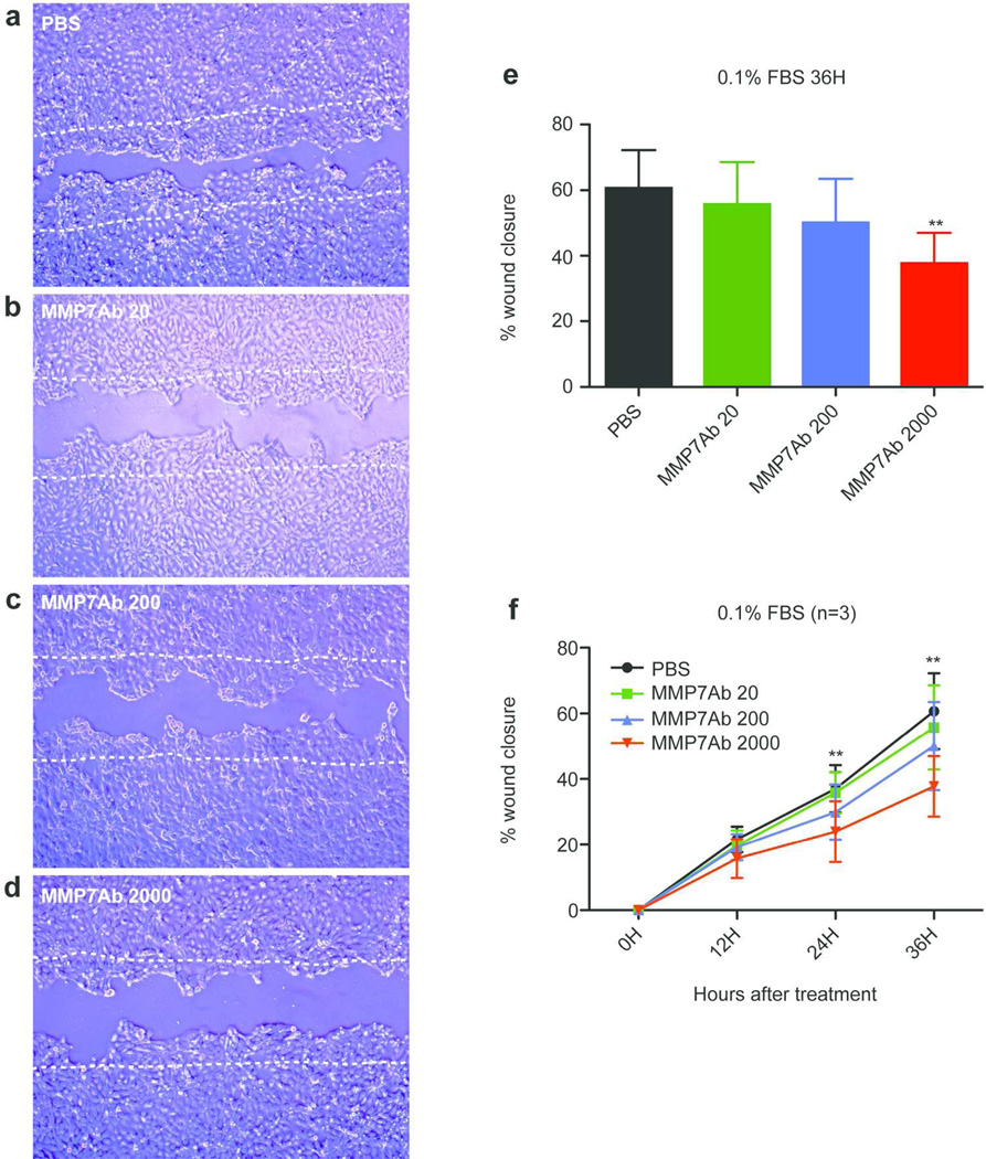
Figure 4. Blocking of MMP7 delayed the migration of A431 cells.
Scratch was made on a culture of A431 cells at 90% confluence and cells were cultured in 0.1%FBS-containing media with or without indicated concentrations of anti-MMP7 antibody (MMP7Ab) for 36 hours. Cells were photographed every 12 hours. (a–d) Representative images of area after a 36 hour cultivation with (a) PBS, (b) 20ng/ml of MMP7Ab, (c) 200ng/ml of MMP7Ab, and (d) 2000ng/ml of MMP7Ab. Two white dotted lines depict the initial area. (e) The bar graph shows mean of percent wound closure for each treatment after 36 hours. (f) The graph depicts the chronological change of the percent wound closure for each condition. An error bar shows S.E.M. (n=3). **; p<0.01 between MMP7Ab2000 vs. PBS and MMP7Ab20 for a 24H treatment, and between MMP7Ab2000 vs. PBS, MMP7Ab20, and MMP7Ab200 for a 36H treatment (repeated measures ANOVA with Tukey’s correction).

