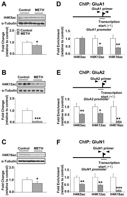
Figure 3.
Chronic exposure to METH promotes hypoacetylation of H4K5, H4K12 and H4K16 on the promoters of AMPA GluA1, GluA2 and NMDA GluN1 subunits. The rats were treated as mentioned in Figure 1. Chronic METH treatment decreases the levels of nuclear (A) H4K5ac; (B) H4K12ac and (C) H4K16ac proteins in the dorsal striatum (n=6 rats per group). Representative photomicrographs show results of three samples per group. For quantification, the signal intensity was normalized to 3-tubulin. ChIP assays (n=6 - 8 rats per group) were carried out using antibodies against histone H4 acetylated at lysine 5 (H4K5ac), at lysine 12 (H4K12ac) and at lysine 16 (H4K16ac) on (D) GluA1, (E) GluA2 and (F) GluN1 DNA sequences. Quantitative PCR was conducted as described in the text using specific ChIP primers directed at GluA1, GluA2 or GluN1 promoters (see Table S2). Values represent means ± SEM of fold changes relative to the controls. Statistical significance was determined by un-paired Student’s t-test. Key to statistics: * p< 0.05; ** p< 0.01; *** p< 0.001 vs. control group.

