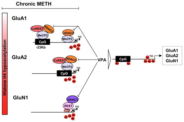
Figure 9.
Schematic models showing chronic METH-induced epigenetic modifications in the dorsal striatum. Under control condition, there exists a balance between histone acetylases (HATs) and histone deacetylases (HDACs) that regulate the histone acetylation/deacetylation status that maintain the baseline transcription levels. However, chronic METH exposure leads to formation of protein repressor complexes MeCP2-CoREST-HDAC2 that cause H4K5, H4K12 and H4K16 hypoacetylation at enhancer or promoter sequences of GluA1 and GuA2 genes. This then leads to decreased expression of these receptors. Rats chronically exposed to METH also show formation of a protein repressor complex that contains REST-HDAC1 that produces hypoacetylation of H4K5, H4K12 and H4K16 at the promoter region of GluN1 and subsequent decreased GluN1 (NR1) expression in the dorsal striatum. Co-treatment of METH-treated animals with valproic acid that inhibits HDAC1 and HDAC2 blocked METH-mediated hypoacetylation and METH-induced decreased expression of these glutamate receptors.

