Figure 1.
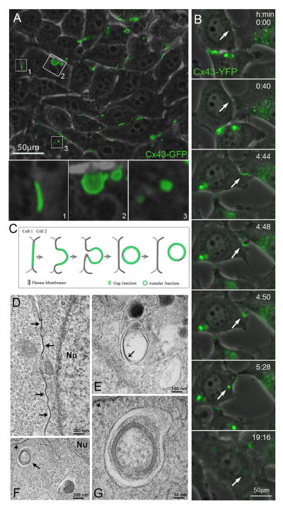
Internalization of gap junction plaques. (A) Combined phase-contrast and fluorescence images of HeLa cells transfected with Cx43-GFP reveal efficient expression and assembly of gap junctions at appositional membranes of transfected cells (visible as green fluorescent lines and puncta such as the one shown in insert (1). Over time, gap junction plaques invaginate into the cytoplasm of one of the two cells (insert 2), detach from the plasma membrane and form endocytosed cytoplasmic annular gap junctions or connexosomes (insert 3). (B) Selected still-images of a time-lapse recording of stably transfected Cx43-YFP expressing HeLa cells showing the formation of a gap junction, its internalization into the cytoplasm of one of the adjacent cells, and subsequent degradation of the generated annular gap junction, indicated by the loss of its fluorescence (marked with arrows). (C) Schematic representation depicting the progressive stages of gap junction internalization shown in A and B. (D) Electron micrograph showing GJs (arrows) at the appositional membranes of mouse embryonic fibroblasts. (E) Electron micrograph showing an internalized GJ structure within a membrane-surrounded compartment, which probably corresponds to a secondary lysosome or autolysosome. Note that the pentalaminar pattern characteristic of GJs is being lost (arrow) in the internalized structure, probably because it is undergoing degradation. (F) Electron micrograph obtained from mouse embryonic fibroblasts starved by incubation in Hank’s balanced salt solution for 1 hour showing a double membrane structure, probably an autophagosome (arrow) that encloses an internalized GJ in close proximity to rough endoplasmic reticulum (short arrow). (G) Electron micrograph showing the internalized GJ enclosed within the autophagosome at higher magnification; the rough endoplasmic reticulum membrane is indicated by the short arrow. Nu: nucleus (A–C reproduced with permission from Landes Bioscience from ref. [91], D–G reproduced with permission from The Company of Biologists from ref. [93]).

