
Fig. 1.
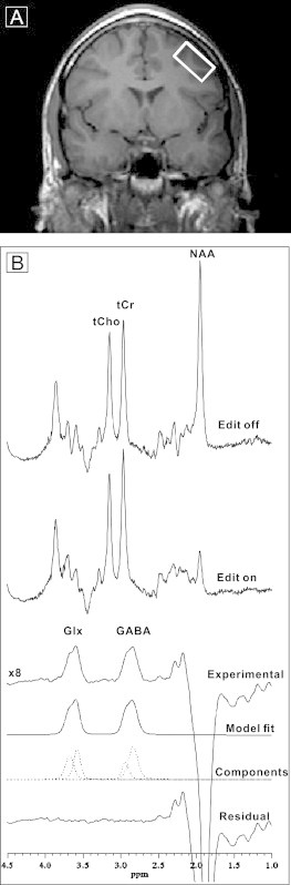
A. Coronal slice through the anterior commissure showing placement of the DLPFC voxel in the left middle frontal gyrus of a patient with schizophrenia. B. An acquisition from this voxel. Six successive curves show spectrum with editing pulse off; editing pulse on; difference spectrum between editing on and off magnified 8-fold showing the resulting Glx and GABA peaks; best fit model spectrum; individual components of the best-fit model; and the residual difference between the measured difference spectrum and the best-fit curve.