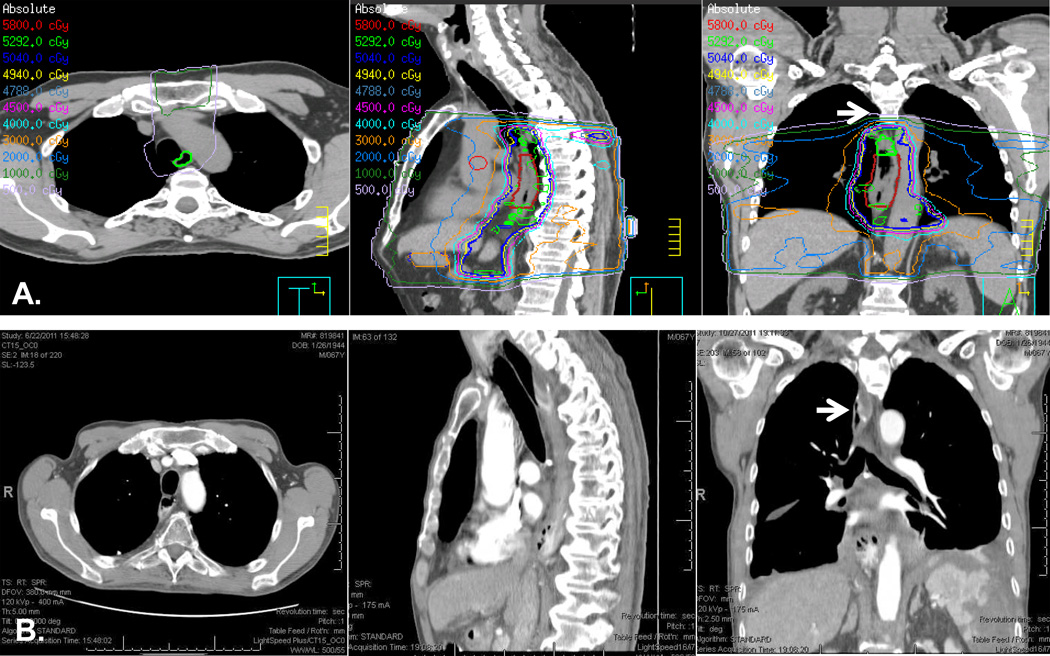
Figure 1. Illustration of a case with out-of-field anastomosis.
A. Simulation CT imaging of a mid-esophageal tumor with treatment plan encompassing areas below the aortic arch. B. Post-Ivor-Lewis esophagectomy CT imaging showing postoperative anatomy. White arrows point to the area of anastomosis above the aortic arch. This patient did not develop anastomotic leak.

