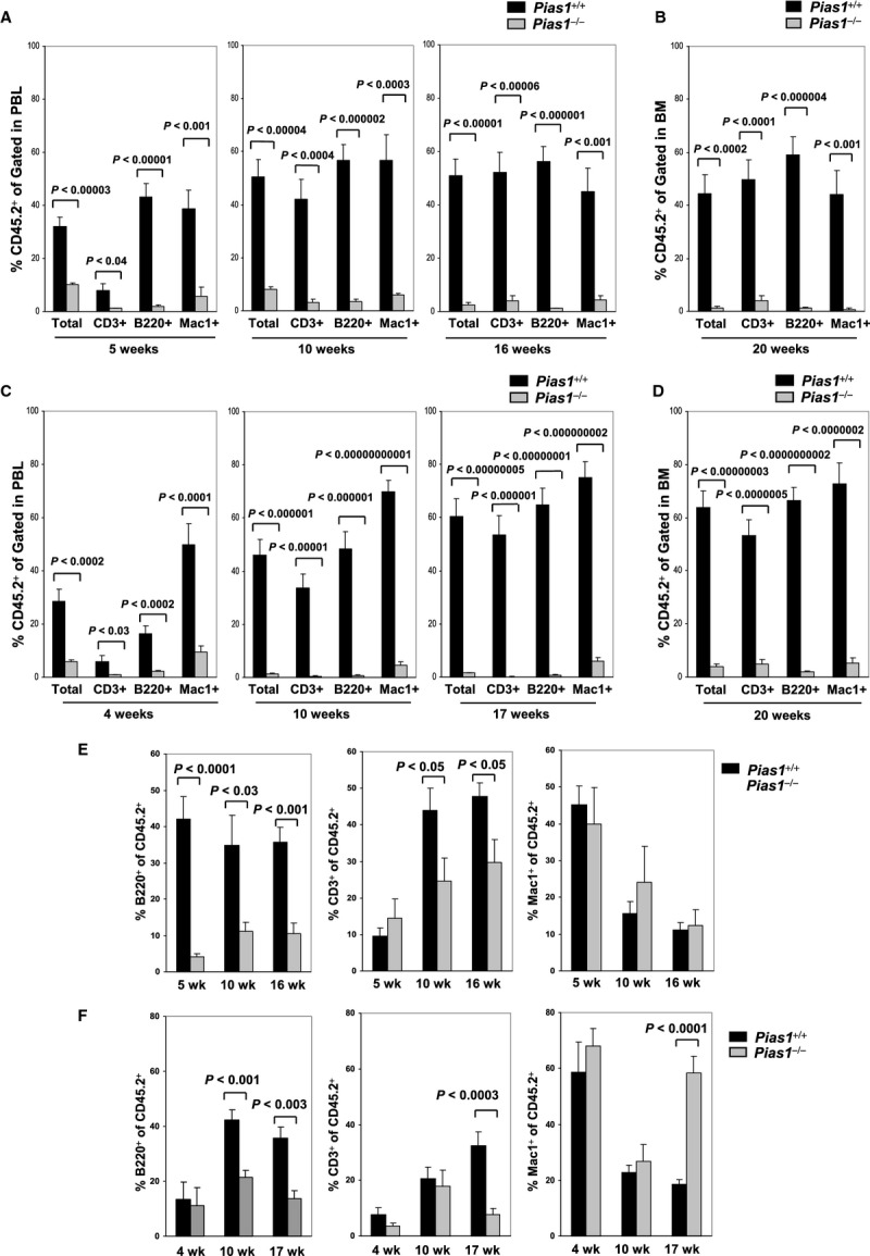
Figure 3.

- In vivo competitive reconstitution assays. Total bone marrow cells (2 × 10⁵) from WT or Pias1−/− littermates (CD45.2+) were mixed with 2 × 10⁵ of WT C57SJL bone marrow cells (CD45.1+) and injected into lethally irradiated WT C57SJL mice. The percentage of T cells (CD3+), B cells (B220+) and myeloid cells (Mac1+) from donor mice in peripheral blood (PBL) were assayed by flow cytometry at 5, 10 and 16 weeks post reconstitution.
- Same as in (A) except that bone marrow (BM) cells from the recipient mice were assayed at 20 weeks post reconstitution.
- Same as in (A) except that FACS-sorted LSK cells (1000) from WT or Pias1−/− littermates were used.
- Same as in (B) except that FACS-sorted LSK cells (1000) from WT or Pias1−/− littermates were used.
- Same as in (A) except that the percentage of T cells (CD3+), B cells (B220+) and myeloid cells (Mac1+) within the donor cells (CD45.2+) in peripheral blood (PBL) were presented.
- Same as in (E) except that FACS-sorted LSK cells (1000) from WT or Pias1−/− littermates were used.
Data information: Shown is a pool of 3 independent experiments in all panels (n = 10). Error bars represent SEM. P-values were determined by non-paired t-test. See also Fig S4.
