Figure 4.

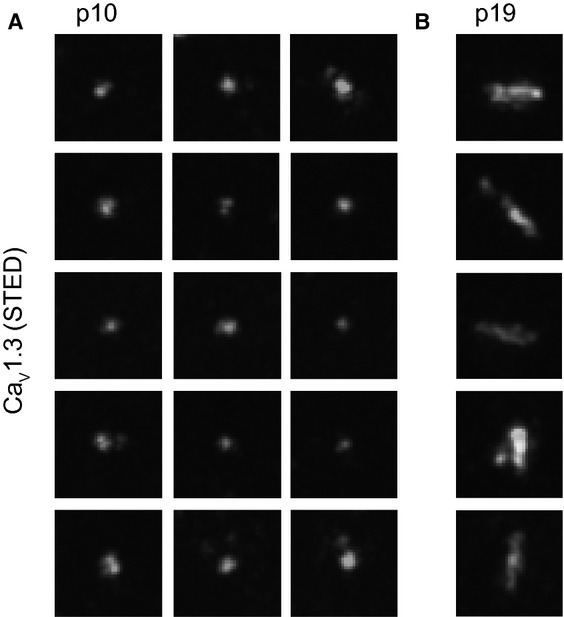
- Projections of STED sections of immunolabeled CaV1.3 clusters in the organ of Corti whole-mount at p10 (0.6 μm step size, total z covered: 4 μm). CaV1.3 clusters in pre-hearing IHCs appeared spot-like (A), in contrast to the stripe-like appearance in mature IHCs (B). Images were processed as in Fig 3G. Pixel size, 20 nm, 700 × 700 nm regions-of-interest centered at individual CaV1.3 clusters.
- Projections of STED sections of immunolabeled CaV1.3 clusters in the organ of Corti whole-mount at p19 (0.6 μm step size, total z covered: 4 μm). CaV1.3 clusters in pre-hearing IHCs appeared spot-like (A), in contrast to the stripe-like appearance in mature IHCs (B). Images were processed as in Fig 3G. Pixel size, 20 nm, 700 × 700 nm regions-of interest centered at individual CaV1.3 clusters.
