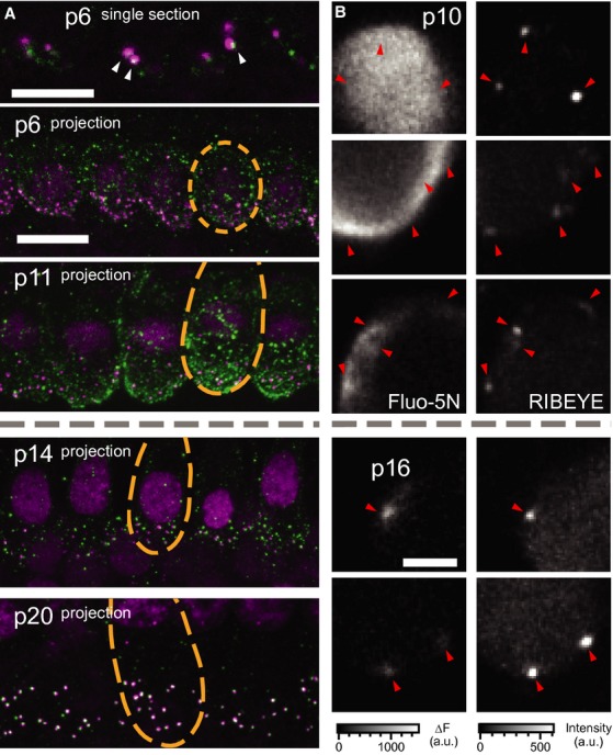
Figure 5.

- Organs of Corti immunolabeled for CaV1.3 (green) and RIBEYE/CtBP2 (magenta). At p6 (top), a single section shows both ribbon-occupied (arrowheads) and ribbonless CaV1.3 spots as well as diffuse membrane staining. At p11 (middle, projection) puncta of CaV1.3 were observed throughout the basolateral membrane, especially the basal pole. By p14 the clusters of CaV1.3 were concentrated in the basal poles near synaptic ribbons, while the entire inner hair cell membrane remained diffusely labeled for CaV1.3. At p20 there was very little extrasynaptic labeling and the cell membrane was no longer diffusely immunoreactive for CaV1.3. Each synaptic ribbon was accompanied by a cluster of CaV1.3 and only few CaV1.3 clusters remained ribbonless. Scale bars, 10 μm.
- Examples of XY scans across the basolateral portion of IHCs before (upper, p10) and after (lower, p14) the onset of hearing. Left and right panels show the increase in Fluo-5N fluorescence during a step depolarization to −7 mV for 254 ms (2 mM [EGTA]i) and peptide-labeled synaptic ribbons (position marked with arrowheads). Note the relatively widespread submembraneous Ca2+ signal in immature IHCs as compared to the local Ca2+ rise in mature IHCs. Still, the highest [Ca2+] was usually observed at the ribbons-occupied AZs. Scale bar, 3 μm.
