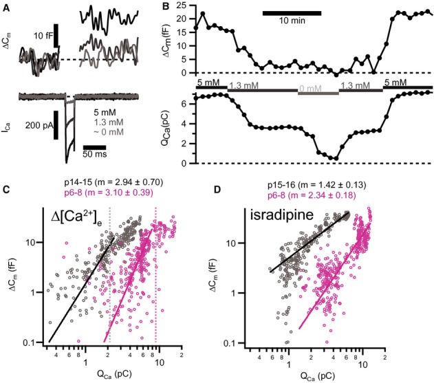
Figure 7.

- Depolarizations (20 ms to −17 mV) elicited inward Ca2+ current (ICa, lower panel) and triggered ΔCm (upper panel) in a representative pre-hearing (p7) IHC. Increasing [Ca2+]e enlarged both ICa and ΔCm.
- Time course for ΔCm and integrated Ca2+ influx (QCa) of the same IHC in (A) for repetitive depolarizations (20 ms to −17 mV) at 60 s interval. Experiment started at 5 mM [Ca2+]e and periods where bath solution was slowly perfused with a different [Ca2+]e are marked by horizontal bars.
- Plotting ΔCm, 20 ms against QCa reveals a supralinear apparent Ca2+ dependence of exocytosis for low QCa. Solid lines represent best fit power functions (ΔCm = A(QCa)m) to the whole data set in this range. Each small symbol represents an individual response to depolarization (p15–16, grey, n = 7 IHCs; p6, magenta, n = 7 IHCs). Dashed vertical lines indicate the average maximal QCa limiting the QCa range used for fitting.
- Decreasing apparent Ca2+ cooperativity of exocytosis through blocking L-type Ca2+ channels with isradipine. Each small symbol represents an individual response to depolarization (p14–15, grey, n = 6 IHCs; p6–8, magenta, n = 7 IHCs).
Data information: In (C) and (D), solid lines show best fit power functions to the whole data set. Average values of the exponent m are indicated on top.
