Figure 1.

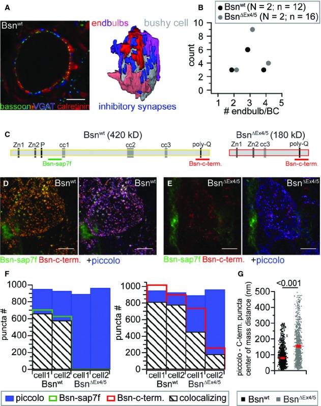
Number of converging endbulbs and bassoon immunolocalization in the AVCN of Bsnwt and BsnΔEx4/5 mice.
A Single confocal section from a stack (left) used for reconstruction of endbulb terminals (right, four endbulbs converging onto the BC in this example), immunolabeled for calretinin (red) as an endbulb marker, bassoon (green) as a marker for AZs and VGAT (blue) as a marker for inhibitory synapses.
B The number of endbulb terminals converging onto a BC remained unchanged in BsnΔEx4/5 mice (N, number of animals; n, number of BCs).
C Domain structure of bassoon and the BsnΔEx4/5 fragment including the epitopes utilized for immunolabeling.
D, E Projection of a confocal image stack labeled for the two bassoon epitopes and piccolo of a Bsnwt (D) and a BsnΔEx4/5 (E) BC.
F Number of puncta and fraction of colocalizing bassoon puncta (left, Bsn-sap7f AB; right Bsn-c-term. AB) with piccolo of two BC for each genotype.
G Center of mass distance between all colocalizing Bsn-c-term. and piccolo puncta of the four cells depicted in (F), illustrating that the BsnΔEx4/5 fragment is not as tightly confined to AZs as wild-type bassoon.
