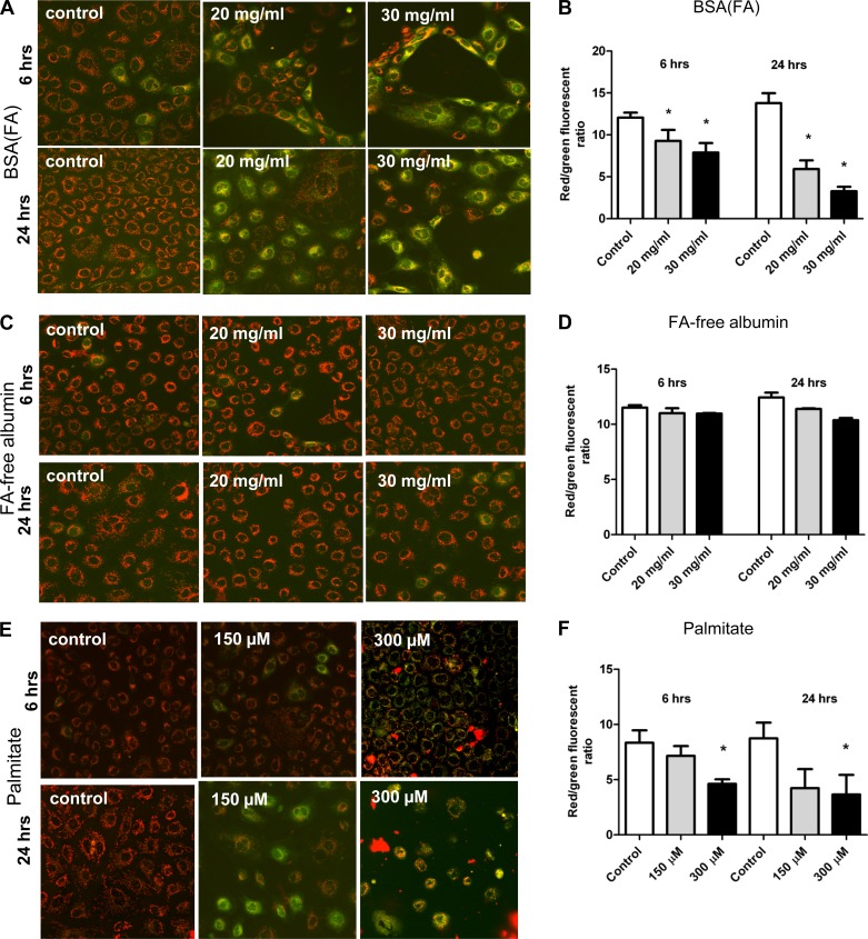
Fig. 2.
Changes in mitochondrial membrane potential (ΔΨ) in NRK-52E cells. Cells were loaded with the cationic JC-1 dye after various exposures to detect changes in ΔΨ by 2 different methods—imaging by confocal microscopy and spectrofluorometric readings by a plate reader. A–B: exposure to BSA(FA) caused loss of ΔΨ as shown by increased diffuse cytosolic green staining and decreased red/green ratios. C–D: FA-free albumin had no effect on ΔΨ as cells maintained dotted red staining of JC-1 and unchanged red/green ratios. E–F: similarly to BSA(FA), palmitate caused a significant loss of potential. *P < 0.05 vs. control, means ± SD; experiments were done a minimum of 3 times.

