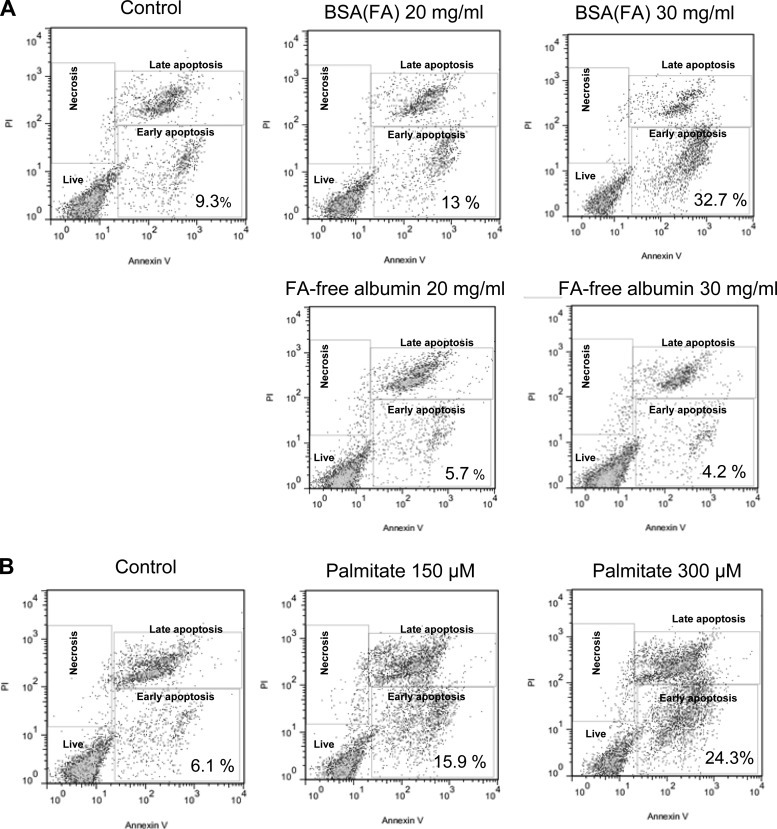
Fig. 4.
Apoptosis of tubular epithelial cells exposed to albumin and FAs. Cells were treated with BSA(FA), FA-free BSA, or palmitate and the population of apoptotic/necrotic cells was analyzed by flow cytometry after an Annexin V/PI assay. A: only BSA(FA) but not FA-free albumin caused an increased ratio of early apoptotic cells after 24 h. B: palmitate treatment also caused a significant increase of the early and late apoptotic populations after 24 h. The flow cytometry results are representative of 3 different experiments, repeated on cells with similar passage numbers.

