
Figure 6.
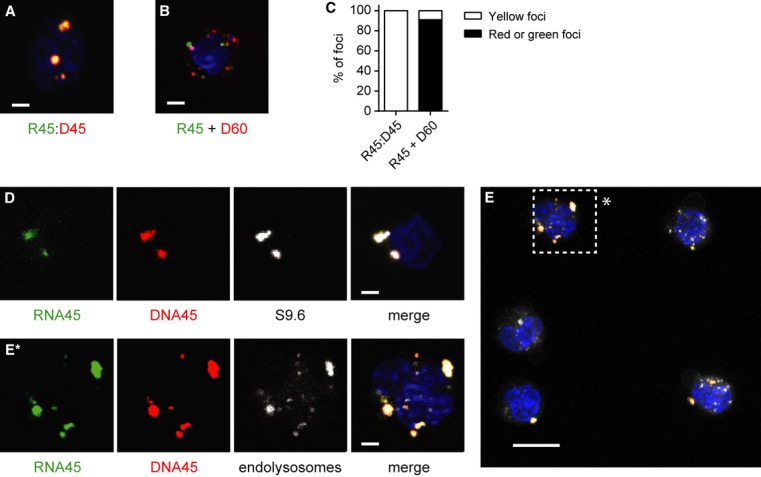
Intact RNA:DNA hybrids are detectable by immunofluorescence inside FLDCs.
- Fluorescently labelled RNA and DNA strands of R:D45 remain equally colocalised in FLDCs following transfection, but non-complementary RNA and DNA oligonucleotides do not.
- Confocal maximum projection image of a cell after transfection with fluorescently labelled R:D45, generated by annealing Cy5-labelled ssRNA45 and Cy3-labelled ssDNA45 oligonucleotides. DNA visualised by DAPI, blue.
- Confocal image of a cell transfected with non-complementary Cy3-ssRNA45 and Cy5-ssDNA60 oligonucleotides.
- Quantification of nucleic acid foci in FLDCs transfected with either fluorescently labelled R:D45 (n = 50) or Cy3-ssRNA45 with Cy5-ssDNA60 (n = 118).
- Intracellular R:D45 is detected by the anti-RNA:DNA hybrid antibody S9.6. FLDCs were transfected with fluorescently labelled R:D45 as for (A), and fixed and permeabilised prior to immunofluorescent detection with the S9.6 antibody. Representative maximum projection confocal image is shown. Scale bar, 2 μm.
- R:D45 frequently colocalises with the endolysosomal marker LysoTracker. Cells were transfected with fluorescently labelled R:D45 as for (A) then incubated with LysoTracker for 1 h. Maximum projection image of a representative field of view. Scale bar, 10 μm.
E* Higher magnification image of a cell (*) in (E). Scale bar, 2 μm.