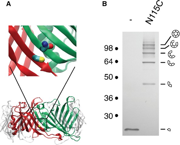
Figure 2.

- Model of the EAEC Hcp1 hexamer. Two adjacent monomers are colored in red and green, respectively. The magnification emphasizes the location of the Cys-38 (red monomer) and Asn-115 (green monomer) residues of the two adjacent monomers.
- Disulfide bond formation between Hcp1 proteins within the hexamer. Cytoplasmic extracts from EAEC Δhcp1 cells producing Hcp1 or Hcp1-N115C after in vivo oxidative treatment were analyzed by 12.5%-acrylamide SDS-PAGE and proteins were immunodetected with the anti-FLAG monoclonal antibody. Positions of the Hcp1 monomer and oligomers are indicated on the right. Molecular weights (in kDa) are indicated on the left.
