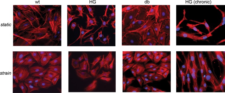
Fig. 3.

Phalloidin staining of static (top) and strained (bottom) samples at acute [high glucose (HG), 24-h culture] and chronic [HG (chronic), 17-day culture] stages. No differences are apparent in cell morphology and cytoskeletal organization between WT and db groups in either static conditions or strain conditions. However, WT cells exposed to high-glucose medium (HG and HG chronic) show altered cell morphology (2nd and 4th columns).
