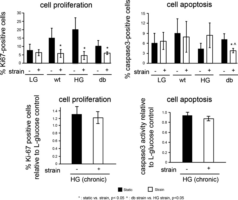
Fig. 5.
Effect of strain on cell proliferation and apoptosis in normal and diabetic cardiac endothelial cells. Ki67 and caspase-3 staining is performed on static controls and cells subjected to 24 h of strain to determine the percentage of proliferating and apoptotic cells. In WT, HG and db experimental groups, cell proliferation was significantly higher in static samples than it was in strained samples (top left). The percentage of apoptotic cells in db group was significantly less under strained conditions compared with static controls. Chronic exposure (17 day) to high-glucose (HG) did not effect endothelial proliferation or apoptosis under either static or strain conditions (bottom).

