Fig. 3.
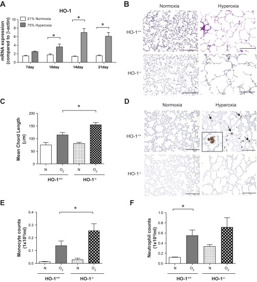
Heme oxygenase (HO-1) knockout exacerbates hypoalveolarization and increases leukocyte infiltration in hyperoxia-exposed neonatal mice. A: whole lung HO-1 mRNA expression from wild-type mice exposed to either normoxia (n = 20–26 mice per group) or hyperoxia (n = 19–23 mice per group) for up to 21 days. Hyperoxic exposure increased mRNA expression, as measured by qPCR (*P < 0.05, ANOVA). B: 3-day-old HO-1+/+ or HO-1-null (HO-1−/−) mice were exposed to air or 75% oxygen for 14 days. Hyperoxia-exposed HO-1−/− mice showed alveolar arrest with large airspaces. Hyperoxia-exposed HO-1-null mice showed exaggerated alveolar simplification compared with wild-type mice. C: group mean chord length data (n = 4–9 per group, *P < 0.05, ANOVA). D: hyperoxia-induced HO-1 expression is observed in lung alveolar macrophages (arrows). Immunohistochemistry was performed with an antibody against murine HO-1 (scale bar = 100 μm). E and F: hyperoxia-induced inflammatory cell influx in neonatal mouse lungs is exacerbated by HO-1 deletion. BALF monocytes (E) and neutrophils (F) were counted 14 days after exposure to room air (21% O2) or hyperoxia (75% O2). Compared with HO-1+/+ mice, HO-1-null mice showed a significant increase in macrophages and a trend toward increased neutrophils (n = 4–7 mice per group, means ± SE, *different from hyperoxia-exposed group, P < 0.05, ANOVA).

