
Fig. 4.
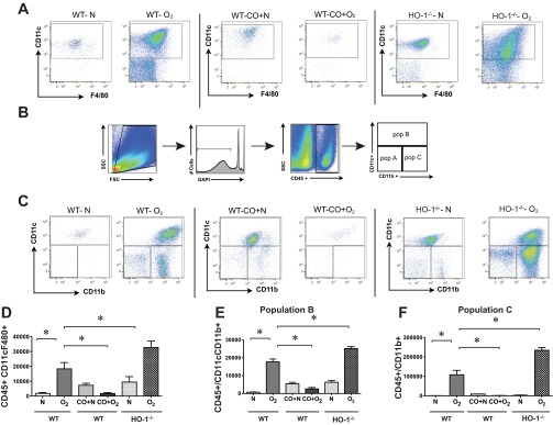
Hyperoxic exposure induces alveolar monocyte/macrophage infiltration into neonatal lungs that is ameliorated by CO-induced treatment. Representative flow cytometric scattergrams of monocyte/macrophage subpopulations within the lungs of wild-type (WT) and HO-1-null mice exposed to normoxia and hyperoxia for 14 days. Wild-type mice were also exposed to normoxia + CO or hyperoxia + CO. Each dot plot is representative of 4 independent experiments. A: CD45+, CD11c+, F4/80+ macrophages. B: gating scheme used in subsequent flow cytometric analysis. C: whole lung live CD45+ cells were analyzed for expression of CD11c and CD11b. D–F: comparison of cell counts for F4/80 and CD11c double-positive macrophages (D), CD11chigh CD11bhigh exudative macrophages (subpopulation B, E), and CD11clowCD11bhigh inflammatory monocytes (subpopulation C, F). Cell counts represent the number of cells per 106 events. Hyperoxia increased the trafficking of macrophage subpopulations into the neonatal lung, and CO treatment attenuated macrophage recruitment. HO-1−/− mice showed increases lung macrophages. n = 4–10 per group, means ± SE, *compared to hyperoxia alone, P < 0.05, ANOVA.