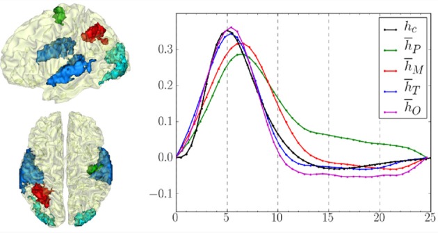
Figure 8.

Left: Definition of regions of interest to investigate hemodynamics variability from JDE-based group-level analysis. Top: Sagittal view. Bottom: Axial/top view. Left parietal area (P) appears in  , left motor area in the pre-central cortex is shown in
  Bilateral temporal regions along auditory cortices and bilateral occipital regions in the visual cortices are shown in
  and
and  respectively. Right: Group-average HRF estimates for the four regions of interest: hP, hM, hT, hO stand for HRF means in parietal, motor, temporal and occipital regions, respectively. hc correspond to the canonical HRF.
