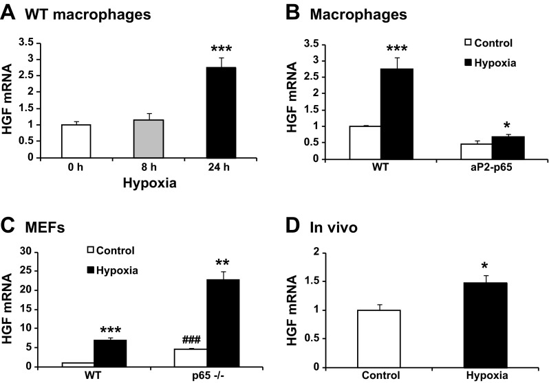
Fig. 5.
Hypoxia induction of HGF expression. A: Hgf mRNA induction by hypoxia. The peritoneal macrophages were exposed to hypoxia (1% oxygen) (n = 3). B: Hgf mRNA in aP2-p65 macrophages. HGF was tested after 24 h hypoxia treatment (n = 3). C: Hgf mRNA in the p65−/− MEFs. The MEFs were exposed to hypoxia for 24 h (n = 3). D: Hgf mRNA in adipose tissue. The Hgf mRNA was determined in epididymal fat of the mice after modest hypoxia (17% O2) treatment for 6 h (n = 5). The results are expressed as mean values ± SE. Compared with 0 h (A) or control (B, C, and D): *P < 0.05, **P < 0.01, and ***P < 0.001; compared with WT control (C): ###P < 0.001.

