Fig. 1.
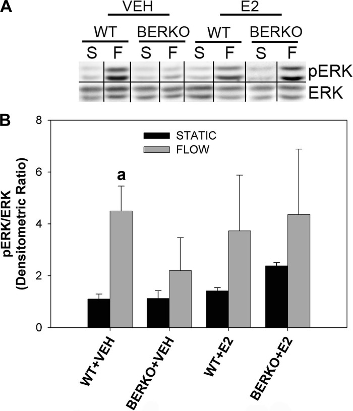
ERK phosphorylation in primary wild-type (WT) and estrogen receptor (ER)β-knockout (BERKO) calvarial cells. A: Western blot analysis of anti-ERK phosphorylation (p-ERK; Y site) and ERK in WT and BERKO cells exposed to static conditions (S) or 30 min of oscillatory fluid flow (OFF) (F) in the absence (VEH) and presence of estradiol (E2). Representative bands from experimental groups are shown. Bands taken from noncontiguous lanes are divided by black lines. B: quantification of p-ERK/ERK densitometric ratios, normalized by the mean of p-ERK/ERK for the WT S VEH group (thus shown as “1”), is presented. Experimental groups contain 2–3 samples. Bars represent normalized means ± SE. Genotype, F, and E2 were not significant factors by a 3-way ANOVA; however, WT + VEH F was significantly greater than WT + VEH S when compared using a Student t-test. aP < 0.05 vs. WT + VEH S.

