Fig. 6.
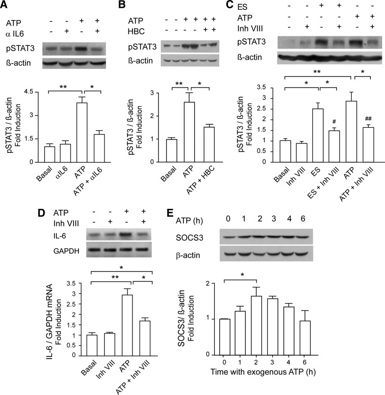
Extracellular ATP activated STAT3 and increased IL-6 expression partially through a positive IL-6 loop. A: blockade of released IL-6 abolishes STAT3 activation evoked by ATP. Myotubes were incubated with 100 μM ATP for 1 h in the absence or presence of an anti-rat IL-6-neutralizing antibody (aIL6, 1 μg/ml, 30 min before and during the ATP stimuli; n = 4). B: ATP induced STAT3 phosphorylation via JAK2. Rat myotubes were incubated overnight with 50 μM HBC or vehicle. Stimulation with 100 μM ATP for 1 h was performed in the presence of HBC. After that, total protein extracts were obtained (n = 3). C: STAT3 inhibitor VIII reduced STAT3 phosphorylation evoked by ES or ATP. Rat myotubes were incubated overnight with 50 μM inhibitor VIII or vehicle; 1 h after ES or 100 μM ATP addition, total protein extracts were obtained, and phosphorylated STAT3 was detected by WB (n = 4). D: STAT3 inhibitor VIII reduced IL-6 expression evoked by ATP. Myotubes were stimulated as in C but processed for total RNA extraction. IL-6 mRNA expression was assessed by conventional semiquantitative RT-PCR, normalized to GAPDH expression, and presented as fold increase of untreated control cells (n = 3). E: ATP evoked expression of suppressor of cytokine signaling 3 (SOCS3), a negative regulator of the JAK/STAT3 pathway. Myotubes were incubated with 100 μM ATP for different times; total protein extracts were obtained, and SOCS3 expression was assessed by WB (n = 4). Values are expressed as means ± SE. *P < 0.05 and **P < 0.01 (as indicated); #P < 0.05 and ##P < 0.01 (compared against condition with only inhibitor VIII), analysis of variance followed by Dunnett's multiple comparison test.

