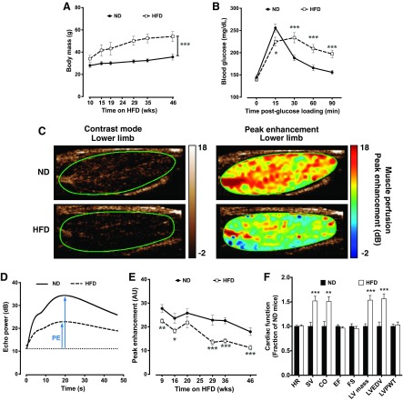
Fig. 1.
Hindlimb skeletal muscle perfusion is reduced in male C57BL/6 mice fed a high-fat diet (HFD). A: body mass. ND, normal diet. B: oral glucose tolerance testing after 46 wk of ND (n = 34) and HFD (n = 33). C: representative contrast-mode ultrasound images of the medial aspect of the lower hindlimb of ND- and HFD mice. D: peak enhancement (PE) is the maximum-minimum video intensity and is an indicator of blood volume. E: average PE in the hindlimb of ND-fed (n = 13–18) and HFD-fed (n = 21–35 except at 20 wk, where n = 12) mice. Only half of the HFD-fed mice were measured at the 20-wk time point. AU, arbitrary units. F: cardiac function in ND-fed (n = 11) and HFD-fed (n = 10) mice. HR, heart rate; SV, stroke volume; CO, cardiac output; EF, ejection fraction; FS, fractional shortening; LV mass, left ventricular (LV) mass; LVEDV, LV end-diastolic volume; LVPWT, LV posterior wall thickness. Data are presented as means ± SE. Data were analyzed with two-tailed, independent t-tests between ND and HFD at each time point. *P < 0.05; **P < 0.01; ***P < 0.001.

