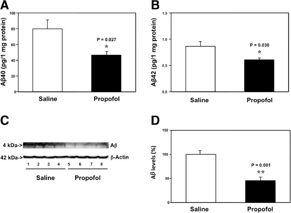
Figure 1.
Propofol decreases Aβ levels in the brain tissues of aged mice. A. ELISA shows that there are lower levels of Aβ40 in the brain tissues of mice following the propofol treatment (black bar) as compared to the mice following the saline treatment (white bar). B. ELISA shows that there are lower levels of Aβ42 in the brain tissues of mice following the propofol treatment (black bar) as compared to the mice following saline treatment (white bar). C. Western blot analysis shows that there are lower levels of Aβ in the brain tissues of mice following propofol treatment (lanes 5 to 8) as compared to the mice following saline treatment (lanes 1 to 4). D. Quantification of the Western blot shows that there are lower levels of Aβ in the brain tissues of mice following propofol treatment (black bar) as compared to the mice following saline treatment (white bar). N = 10.

