Figure 1.

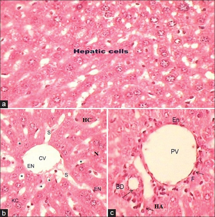
(a-c) Photomicrograph of transverse section of liver (H and E, ×40) of untreated Group I showing the normal hepatic cells, thin walled central vein, hepatic sinusoids (S; *), portal vein, hepatic artery and bile ducts

(a-c) Photomicrograph of transverse section of liver (H and E, ×40) of untreated Group I showing the normal hepatic cells, thin walled central vein, hepatic sinusoids (S; *), portal vein, hepatic artery and bile ducts