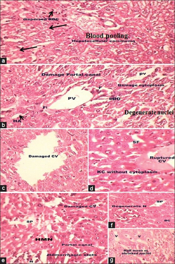
Figure 2.

(a-g) Photomicrographs of transverse section of liver of N-nitrosodiethylamine-intoxicated mice (H and E, ×40) showing the distorted liver architecture and arrow indicated tumor cells contain intracytoplasmic violaceous, hyaline globules that represents proteins produced by the tumor cells (a). Damaged portal vein, hepatic artery and bile ducts (b), ruptured central vein (c and d), degenerate nuclei (N; f) with damage hyperchromatic malignant nuclei (HMN; e) and vacuoles (V; g) were also observed
