Figure 3.

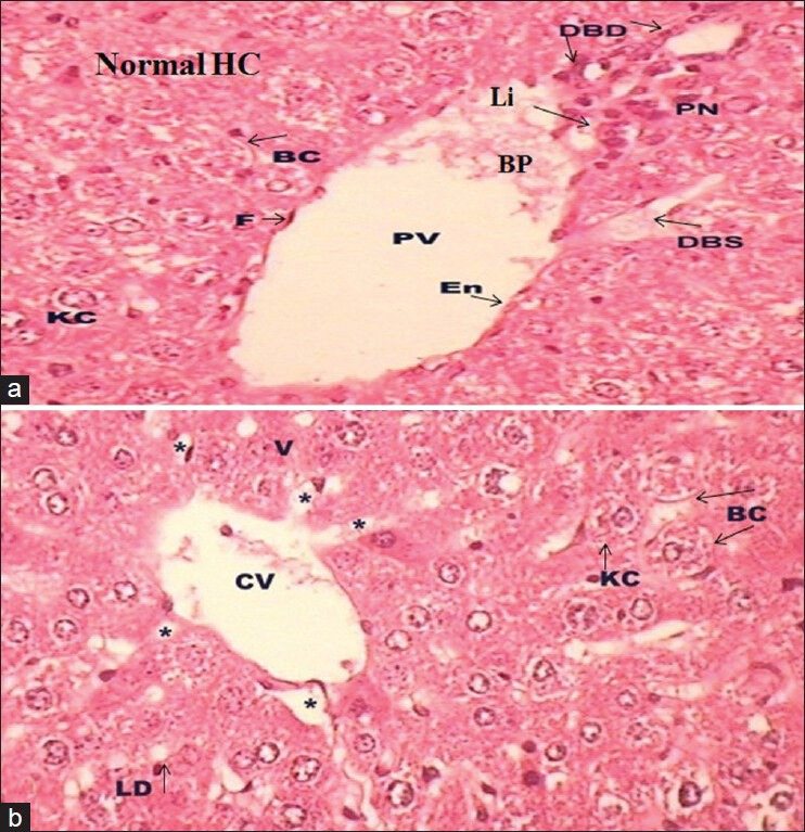
(a-b) Photomicrograph of transverse section of liver (H and E, ×40) of Group III (a) and Group IV (b) showing normal architecture with some centrilobular swelling (a) and some lipid droplets (LD; b)

(a-b) Photomicrograph of transverse section of liver (H and E, ×40) of Group III (a) and Group IV (b) showing normal architecture with some centrilobular swelling (a) and some lipid droplets (LD; b)